No products.
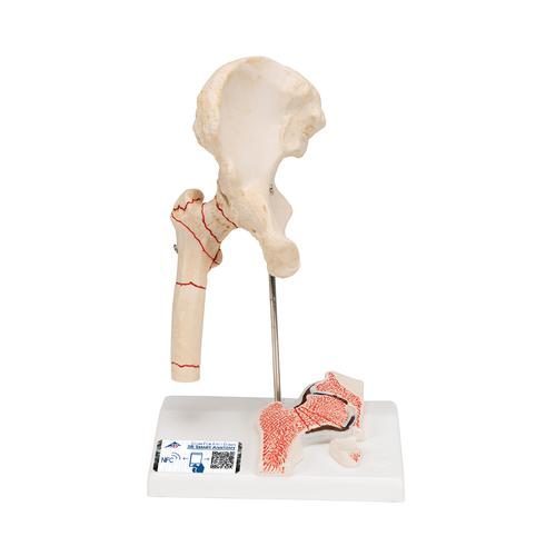
Human Femoral Fracture & Hip Osteoarthritis Model – 3B Smart Anatomy
£99
Delivery:
9-13 working days
9-13 working days
Out of stock
When ordering an item that is temporarily out of stock, your order is noted as remaining and sent out as soon as it is available again. Backlogged orders are handled in turn.
Reviews
There are no reviews yet.